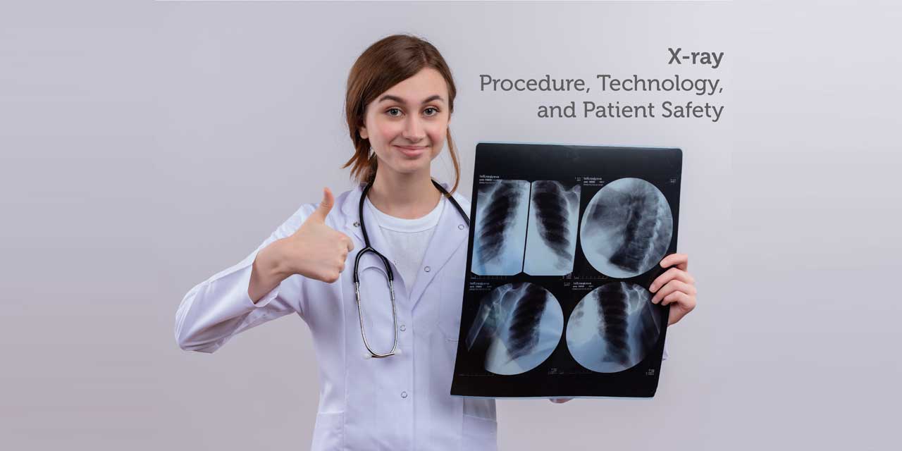
Introduction
Medical imaging techniques such as X-rays, magnetic resonance, and ultrasound, allow the physician to see inside the body of the patient to identify and diagnose the medical problem or disease.
What are X-rays?
X-rays are an energetic form of electromagnetic radiation that is used to reveal the images of the bones present beneath the skin. The radiation produced by the X-ray (radiograph) can penetrate the soft body tissues and not the bone and produces shadow images on the photographic plates. The dense material likes bones and metal shows up as white on the X-ray result plate.
If the X-rays pass through fat and muscles, they appear gray, as they absorb less. The air shows the black color as it absorbs the least. A small dose of X-rays ionizing radiations are used to produce images of the bones, and are most frequently used for medical imaging. The advanced focused X-ray beams are used for the testing application, imaging bones, identifying cracks in the structural components, and several nondestructive evaluations. X-rays can be classified into soft and hard X-rays, and the difference in them is based on their source.
Painless and Quick Test
X-ray is a painless and quick test, in which a patient is positioned, and the target body structure is located between the X-ray detector and X-ray source. The X-ray beams pass through the body tissues and are absorbed depending upon the radiological density (determined by both atomic number and the density) of the material they pass through. The bone calcium atomic number is higher, and this property helps the bones to absorb X-rays readily, and they produce high contrast on the X-ray detector. Hence, in a radiograph, the bones structure appears white, and other tissues present a black background.
Benefits of X-ray test:
- Fast and easy imaging test.
- Helps the physician to view and assess bone injuries, bone dislocation, and bone fracture.
- It is particularly used in emergency diagnosis and treatment.
- Locate foreign objects in bones or in soft tissues.
- It clearly shows up the fractures and infection in the teeth and bones.
X-ray test also helps to detect various diseases and bodily defects:
- Osteoporosis (measure bone density)
- Enlarged heart (a sign of heart failure)
- Breast cancer (mammography)
- Reveal bone tumors
- Detects abnormal bone growth and changes in bone
- Dental decay
- Arthritis
- Blocked blood vessels
- Digestive tract problems
- Chest X-ray helps to detect lung infections, lung cancer, tuberculosis, and pneumonia.
X-ray procedure:
Step 1. X-ray examination does not require any preparation. Before the process, you may be asked to wear a gown, remove eye lenses, metal objects, jewelry, dental appliances, as these may interfere with the X-ray results.
Step 2. In many cases, a liquid called contrast material (iodine or barium) is given before the test (received via swallowing or enema, or injection). It helps in highlighting a specific area during the imaging test.
Step 3. The technician positions the body to obtain the desired views, and one must stay still during the test to prevent the formation of blurry images. The pelvic area and breasts are protected from radiation using a lead apron.
Step 4. The X-ray device produces a small burst of radiation that passes through the body. The patient will also be asked to reposition their body for another view and to obtain a view from different angles.
Step 5. Follow-up exams may be needed to see if the treatment is working and also to detect the change in an abnormality over time.
Result:
The results of X-rays can be seen on the X-ray films and computers within few minutes and are stored electronically. This helps the doctors to diagnose the disease easily and helps in disease management. The results are further obtained in a photographic film or a special detector.
Which is the best X-ray imaging technology available?
Trivitron Healthcare portfolio of radiology products includesFixed and Mobile Radiography Systems in both; Analog and Digital variants, series surgical C-Arm systems available in 3.5kW and 5.0kW power output with Image Intensifiers & Flat Panel Detectors, a comprehensive range of Radiation Protection Products, Anti-scatter x-ray grids and Imaging Accessories including Computed Radiography systems, Dry Films for DICOM printers, Screen Films, Analogue Cassettes and Screens.
For more information visit https://www.trivitron.com/products/radiography–c-arm-systems.
Risks associated with X-ray imaging test:
The amount of radiation released during an X-ray procedure depends on the target organ been examined. The radiation exposure from X-ray is low, and the accurate diagnosis and benefits of the tests are more compared to the risks.
Patient safety:
* One can resume their normal routine after the X-ray test. If during the test, you have received the contrast medium drink plenty of water to get rid of it from the body.
* The patient must inform their technician and physician if they are pregnant. Many imaging tests are not performed during pregnancy as these radiations can cause damage to the fetus. If the test is necessary, then precautions will be taken to minimize radiation exposure to the fetus. Many a time, the healthcare professional decides to go for another imaging method that does not use radiation or ultrasound scan. If a pregnant woman goes for an X-ray (unless necessary), then there is an increased risk that the baby can develop cancer in childhood.
* Modern X-rays produce controlled beams to minimize scatter radiation. This ensures that the patient’s body parts that are not being diagnosed are at minimal exposure to radiation.

