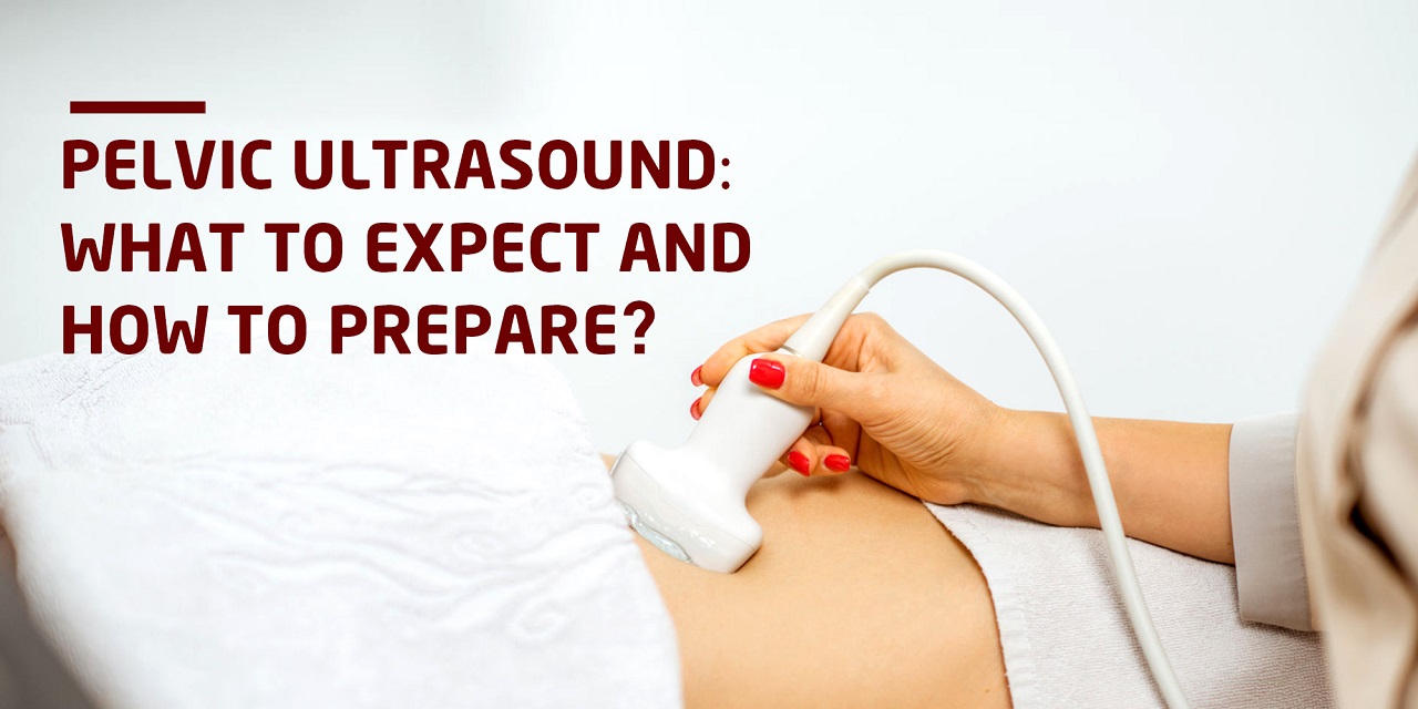
If the physician has advised a pelvic ultrasound, this indicates that the physician is interested in assessing the patient’s uterus, ovaries, and bladder for any problem or abnormality. Ultrasound, unlike other diagnostic technologies, utilizes sound waves rather than radiation to create pictures of the human body. It is especially beneficial for examining soft tissues, organs, and fluids. A pelvic ultrasound typically includes two components: the transabdominal examination and the transvaginal examination. Understanding the exam’s aim and the preparation and method for the first section will help one navigate the procedure smoothly.
The transabdominal ultrasound exam offers a “large view” of the pelvic organs, including their size, shape, position, and orientation, as well as any anomalies. As the term suggests, sound waves are sent through the patient’s lower abdomen to provide a comprehensive image of the uterus, ovaries, and surrounding tissue. A full bladder permits the sound waves to flow into the abdominal cavity without losing substantial power, resulting in a more distinct image.
As a full bladder improves the transabdominal pelvic picture, the patient must ensure that their bladder is full prior to this examination. Normal bladder preparation for this procedure is consuming three 8-ounce glasses of water 45 to 60 minutes before the evaluation. This quantity is sufficient for well-hydrated individuals to generate a full bladder. If one uses caffeine regularly, this quantity of water may not be sufficient to fill the bladder. As a diuretic, caffeine causes cells to lose water. If one uses caffeine regularly without drinking enough water, they may become so chronically dehydrated that 24 ounces of water will be absorbed by the tissues before reaching the bladder.
Therefore, in such a case in caffeine user’s bladder may not be sufficiently full before the ultrasound scan, necessitating extra water and additional time for the bladder to fill. To prevent this, one must hydrate their body the day before the test by consuming eight 8-ounce glasses of water and emptying the bladder as necessary. This strategy flushes the system with much-needed water so that on the day of the test, the body tissues are well-hydrated and the 24 ounces of water one consume will move directly to the bladder within the given time.
Before the test, the technician will apply gel to the lower belly area to begin the examination. Then, the technician will pass a transducer through the gel to transmit sound waves into the body. These sound waves pass into the pelvic cavity, impact the uterus and ovaries, then return to the transducer to produce an image on the screen. Only the pressure of a full bladder will be noticeable. The technician will photograph and measure the pelvic organs, noting any abnormalities. Typically, the whole treatment takes less than twenty minutes, following which one will be permitted to use the restroom. If the physician has ordered a transvaginal examination, the technician will next do the transvaginal portion of the pelvic exam.
A transabdominal ultrasound is a rapid, painless, and radiation-free diagnostic technique that the physician may use to get more information about the patient’s pelvic organs. This test does not offer any difficulty even if the patient keeps their bladder full for many minutes.

