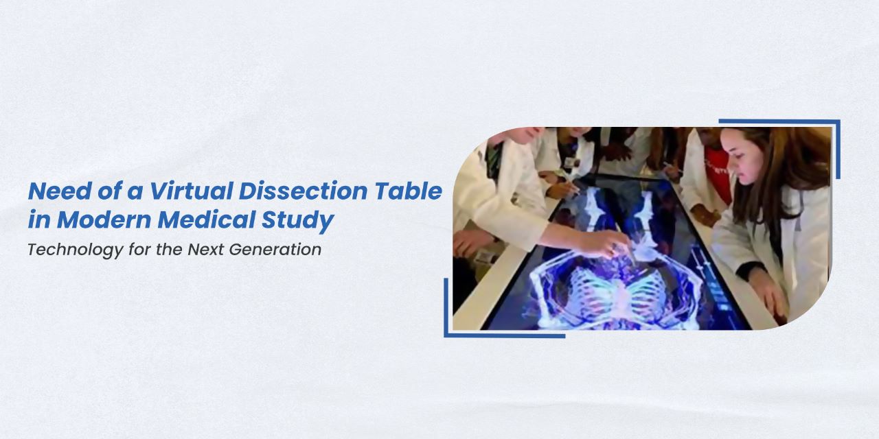
The need for advanced and innovative teaching tools has become increasingly important in modern medical education. One such tool that has gained popularity in recent years is the virtual dissection table. A virtual dissection table is a high-tech device that uses advanced imaging technology to create a detailed, interactive, and 3D digital representation of the human body which medical students can manipulate and explore.
Traditionally, medical students learn anatomy through the dissection of cadavers. However, this approach has several limitations, including limited access to corpses, ethical concerns and practical issues such as space availability and proper ventilation.
Virtual dissection tables have become integral to modern medical education due to their numerous benefits over traditional methods. It offers a range of benefits over traditional dissection methods. One significant advantage is that it eliminates the need for physical cadavers, which can be costly, difficult to obtain and pose health risks to students. Instead, students can use the virtual dissection table to study anatomy and physiology in a safe and controlled environment.
These modern dissection virtual tables offer a solution to these problems by providing an alternative method of learning anatomy that is accessible, safe and effective.
It allows students to manipulate and dissect virtual 3D models of the human body using a computer and specialized software. This technology provides students with a realistic, interactive, and dynamic learning experience that closely mimics the actual dissection process. Students can view different layers of the body, rotate and zoom in on specific structures and even perform virtual dissections.
It enables students to study anatomy at their own pace, review complex concepts repeatedly, and practice procedures without the risk of damaging the specimen. It also supports students to learn anatomy more efficiently and cost-effectively as they do not require the same level of resources and maintenance as cadavers.
Moreover, virtual dissection tables can facilitate collaboration and communication between students and instructors. Students can collaborate on virtual dissections, share their findings, and receive real-time instructor feedback. This technology also enables instructors to create customized lessons, simulations and assessments to enhance student engagement and learning outcomes.
With its high-resolution images and interactive features, students can explore the human body in previously impossible ways. They can zoom in on specific organs or body parts, rotate images in any direction, and even simulate surgeries or medical procedures.
Another benefit of the virtual dissection table is that it can be used for collaborative learning. Multiple students can work together on the same image, sharing real-time insights and observations. This can help students develop teamwork and communication skills, which are essential in the medical field.
A virtual dissection table is an innovative tool changing how medical students learn about the human body. Its many benefits, including safety, cost-effectiveness, and interactivity, make it a valuable addition to modern medical education. As technology evolves, virtual dissection tables will likely become an increasingly common sight in medical schools and training programs worldwide.

