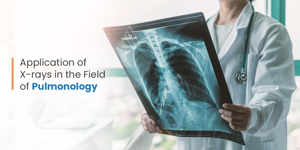
Pulmonology is a branch of internal medicine that deals with diseases related to the respiratory system. A pulmonologist is recommended when one suffers from long-term respiratory conditions like asthma, tuberculosis, lung cancer, and more.
X-ray imaging plays a significant role in the diagnosis and treatment of respiratory conditions. The electromagnetic ionizing radiation from the X-ray machine produces a greyscale picture of the lungs, heart, ribs, and spine to determine the illness. A chest X-ray enables the identification of the following lung conditions:
- Emphysema
- Pneumonia
- Lung Cancer
- Cystic Fibrosis
- Tumors that are on the lungs or near the lungs
- Bronchitis
- Lung congestion due to congestive heart failure
- Fluid or air around the lungs
- Heart failure or other heart-related problems &
- Medical conditions related to spine and ribs
What happens during a chest X-ray?
During a chest X-ray, the patients are requested to wear a lightweight X-ray gown and remove metallic objects like jewellery from their neck/chest areas as the objects can block the penetration of radiation, thereby making the image unclear and less accurate. The imaging procedure also requires the patients to hold their breath as inflated lungs increase the visibility of different tissues within the chest area.
The patients will be asked to move their arms and shoulders into a variety of postures and angles for interpreting all or specific sides of the chest. The duration of an X-ray screening ranges from 5 to 15 minutes and is performed by a well-trained technician or a Radiologist
Trivitron’s Contribution to Pulmonology:
Generally, chest X-ray equipment consists of a wall-mounted, box-like device storing the X-ray film or a detector that captures the picture digitally. Alternately, the X-ray tube may be suspended above a patient-supporting table. The X-ray film or digital detector is kept in a slot under the table.
The portable X-ray machines are equipped to be transported to the hospital bed or emergency room of the patient. A flexible arm is linked to the x-ray tube. The technician extends the arm over the patient and positions the X-ray film holder or image recording plate underneath the patient.
Trivitron’s range of digital X-ray equipment geared with environment-friendly features can be used in nursing homes, home care imaging, small and large animal veterinary, and military field operations too.
Conclusion:
X-ray imaging is beneficial for emergency diagnosis and treatment due to its simplicity, speed, and ease of use. The equipment also leaves no adverse impact on the body when the radiation is within the range of the diagnostic test. To conclude, Trivitron Healthcare offers a range of portable, reliable, and safe X-ray imaging equipment such as surgical C-Arms, X-Ray RAD Room and Mobile X-Rays, and many others. To know more click here

