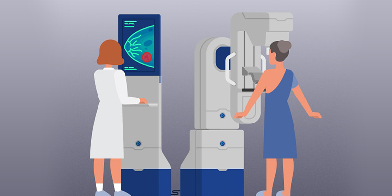
Developments in mammography imaging have changed the manner in which radiologists and oncologists work, as well as, the lives of the ones who have experienced this essential technology. Advancements in mammography imaging technology are reportedly already positively impacting the health of women of all ages in real ways that improve care delivery and outcomes; they include an increased and improved ability to detect breast cancer at the earliest possible moments, confirmation of the benefits of 3D imaging for early breast cancer detection, and the ability of artificial intelligence (AI) to accurately and reliably classify breast tissue thickness without the inherent human inconstancies.
Digital mammography marked a new era of improved cancer detection
The transition to digital mammography from film mammography, has been a turning point in the detection and diagnosis of breast cancer malignancies. Mammogram technology making use of the digital 2D imaging has been widely accepted standard method for breast cancer screening and detection because of several improvements over screen film mammography. For instance, 2D mammography yields better quality visualizations to detect calcification and observe through denser breast tissue for cancer confirmation. Also, digital images give the flexibility of editing and fine-tuning to enhance the image quality, thus allowing radiologists to get a better visuals.
Digital mammography, particularly 2D imaging has proven to be greatly beneficial for women aged 45-70 as findings reveal 2D mammography has translated into a 14 percent higher rate of detection for invasive early-stage grade one and grade two breast cancers.
Further to this, another significant benefit of 2D mammography is, despite the increase in detection rate, this technology has led to a significant decrease in recall rate for false positives, meaning patients need not to undergo unnecessary further screening because of abnormal results.
3D mammography enabled early cancer detection in older women
The integration of 3D imaging techniques in mammography proved to be greatly effective in detecting breast malignancies. Ongoing studies have confirmed several advantages of 3D methods such as a reduction in false positives and abnormal interpretations that lead to unnecessary additional screening and worry. The combination of tomosynthesis in breast mammography has also resulted in greater accuracy in the prediction of the likelihood that patients who received a positive screening result would be diagnosed with breast cancer, later. Tomsynthesis also resulted in higher specificity than exams conducted with 2D mammography, meaning there were fewer false positives.
Additionally, researchers concluded that fewer lymph node-positive cancers were detected in tomosynthesis exams because the presence of cancer was being found at an earlier stage, the ultimate goal of mammography.
AI driven algorithms help in categorizing dense tissue for mammograms
Women with dense breast tissue are at significantly greater risk for developing cancer. Research scientists have made use of artificial intelligence to build an algorithm that can quickly and accurately assess breast density. Today, a number of mammography procedure rely on this algorithms and almost every digital mammogram is automatically assigned a density rating before a radiologist’s evaluation thus standardizing and automating routine breast tissue density assessment
Various research are still underway to further upgrade the mammography technology, however, there is still much in the feasibility stage of development, these techniques are proven to be useful additions to cancer care for particular groups of women.

