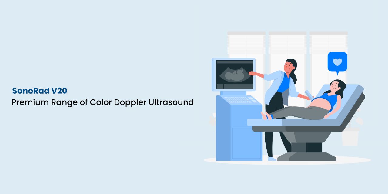
SonoRad V20 Color Doppler Ultrasound Machine uses advanced technology to produce accurate and detailed images of organs and tissues in the body.
It is equipped with various advanced technologies to produce high-quality images, including digital signal processing, harmonic imaging, and tissue Doppler imaging. These technologies work together to produce images with excellent resolution and detail, making them an ideal choice for various diagnostic applications.
The machine comes with a range of probes, including convex, linear, and phased array probes, and its ergonomic design and easy to use, making the user comfortable for long scanning sessions.
One of the major benefits of the SonoRad V20 is its ability to produce 3D and 4D images, which provide a more detailed view of the anatomy being scanned. This feature is particularly useful for obstetric and gynecological applications, where it can provide valuable information about the health of the fetus and the reproductive system.
Another advantage of the SonoRad V20 is its high frame rate, which allows for real-time imaging of moving organs and tissues. This feature is particularly useful in cardiac applications, where it can provide detailed information about the heart’s function.
Technology
Pulse Inverse Harmonic Imaging (PIHI) is a technique used in SonoRad V20 that enhances image quality by reducing clutter and noise. It achieves this by transmitting pulses at a higher frequency than the fundamental frequency of the transducer, causing harmonic waves to be produced. These harmonic waves are then used to create an image, which has improved contrast resolution and reduced noise.
Speckle Reduction Imaging (LanSRI) is a technique that uses sophisticated algorithms to reduce speckle noise, which is a common artifact in ultrasound imaging caused by interference patterns from scattered sound waves. The result is a smoother and more visually pleasing image.
Multiple Compounding Imaging (LanBeam) is used in color Doppler ultrasound to reduce shadowing effects and enhance image quality by combining multiple ultrasound images obtained from different angles. The technique is useful for visualizing complex anatomy and pathologies.
These techniques utilized in SonoRad V20 help enhance image quality, improve diagnostic accuracy, and make the examination more efficient and effective.
Clinical Features
Color Flow Peak Velocity Capture help measures the highest peak velocity in a blood vessel. It is useful in diagnosing stenosis and occlusion in arteries and veins.
Tissue Doppler Imaging (TDI) measures the velocity of tissue movement. It is useful in evaluating cardiac function and detecting abnormalities in myocardial contraction.
Real-time 3D Imaging (4D Imaging) allows the visualization of a three-dimensional image in real time. It is useful in obstetrics and gynecology for evaluating fetal anatomy and cardiac function.
Auto Follicle automatically measures and tracks the growth of follicles in the ovaries. It is useful in monitoring ovulation and fertility treatment.
Triplex Mode combines B-mode, color Doppler, and spectral Doppler imaging into a single display. It is useful in evaluating blood flow and tissue perfusion in real time.
Auto OB calculates fetal measurements and estimates gestational age. It is useful in obstetrics for monitoring fetal growth and development.
Auto IMT measures the thickness of the intima-media complex in the carotid arteries. It is useful in assessing the risk of cardiovascular disease.
Panoramic Imaging in SonoRad V20 allows the visualization of a large area of anatomy by stitching together multiple ultrasound images. It is useful in evaluating organs with a wide field of view, such as the liver and spleen.
Tomographic Ultrasound Imaging helps visualize a section or slice of anatomy. It is useful in evaluating the depth and extent of lesions and masses.
Nodule in Thyroid detects and measures nodules in the thyroid gland and is useful in evaluating thyroid nodules and detecting thyroid cancer.
Real-time Doppler Auto-traces the Doppler waveform and calculates parameters such as peak velocity and resistance index. It is useful in evaluating blood flow in real time.
Multi-frequency Imaging allows multiple frequencies to visualize different depths of anatomy. It is useful in evaluating structures with varying depths, such as the pancreas and kidneys.
Trapezoid Imaging offers a wider field of view by projecting a trapezoidal image onto the screen. It is useful in evaluating structures with a wide field of view, such as the fetal heart.
Duplex and Triplex Modes are features in ultrasound machines that combine B-mode imaging with either spectral Doppler (Duplex) or color Doppler (Triplex) imaging. They are useful in evaluating blood flow and tissue perfusion.
PW, CW & HPRF help measure blood flow velocity using spectral Doppler ultrasound. They are useful in evaluating blood flow in different types of vessels, such as large arteries and veins.
Real Skin Imaging is a feature in SonoRad V20 that allows for the visualization of skin layers. It is useful in evaluating skin lesions and detecting skin cancer.
12 Times Zoom allows for the magnification of the image up to 12 times its original size and is useful in evaluating small structures and lesions.
Variable Doppler gate cursor from 0.5 mm-20 mm is a feature in ultrasound machines that allows for adjusting the size of the Doppler gate cursor. It is useful in evaluating blood flow in vessels of different sizes.
Auto Angle Correction is a feature in ultrasound machines that automatically adjusts the angle of the Doppler beam to align with the direction of blood flow. It is useful in obtaining accurate measurements of blood flow velocity.
Extraordinary Ergonomics
This product boasts extraordinary ergonomic features, such as a 19-inch LED monitor with an articulating arm, an intuitive and height-adjustable control panel, storage space, three probe ports, a one-step caster, a lock mechanism, and a four-wheel swivel, allowing for a wide viewing angle of up to 170 degrees. Additionally, it comes equipped with an omnidirectional mechanical arm, lighting for the keyboard controls, and a high-quality stereo audio speaker system with input and output connections on the rear panel.
Designed to enhance user-friendly workflow, it offers extensive ultrasound functions, including an intuitive touch panel operation, the ability to measure a projected image via the touch panel, zoom in/out on the projected image, and rotate or erase on a projected 3D/4D image on the touch panel. Users can also define their gestures using two fingers for even more functions.
SonoRad V20 is a unique color doppler ultrasound imaging machine that offers a range of advanced features and capabilities. Its advanced technology, probes, and imaging capabilities make it ideal for various diagnostic applications, including obstetric and gynecological, cardiac, and abdominal imaging.

