Introduction
If we talk about the major technology available at ambulatory surgery centers or hospital surgical departments, then the question arises what to consider a Flat-panel Detector (FPD) or an Image Intensifier?
Armed with the right information, provided in this informative article one can choose the best technology that would give an optimum return on investment. The first C-arm was introduced in the year 1950 and developed using existing X-ray technologies and it has revolutionized the area of modern medicine. The unique design of the C-arm helps medical practitioners by offering real-time monitoring and making the necessary corrections to achieve better outcomes.
The C-arm is made up of two components, an X-ray generator, and an image intensifier or flat-panel detector to produce an image. It includes other components like a moveable C-arm and a workstation used to view, store and manipulate images. Generators help in the production of X-rays and detectors or intensifiers absorb and concert these X-rays into an image that is to be displayed on the monitor.
Key Features of Each Technology:
Technology Used
Image intensifier was developed using analog technology and works by converting X-rays into photons for viewing the images. To achieve this the X-rays are first converted into the photons of light as they enter into input phosphor, and these are converted into electrons with the help of a photocathode. The focusing electrodes further amplify these electrons and the outer phosphor converts the amplified electrons back into photons for viewing that is captured by a camera and sent to a monitor for display.
Flat-panel detectors use digital technology and produce a clear image, and these detectors convert X-rays directly into digital data. It also eliminates the need for an optical conversion step and produces an image with less distortion.
Degradation and Distortion of Image
Flat-panel detectors show minimal image degradation over a longer period of time as compared to the image intensifiers. It also has a flat-panel design, which eliminates the geometric distortion. The image intensifiers C-arms provide high-quality images for several years, but the image gain and quality of the image may decrease over time. The presence of input phosphor also produces a peripheral field of view distortion.
Quality Images
The flat-panel detectors are sensitive compared to image intensifiers. They provide patients with lower radiation doses while imaging along with it offers higher and more consistent image quality.
Magnification
In a flat-panel detector, magnification is achieved without reducing the scale. Image intensifiers for magnification utilize collimation, a narrow beam produces a detailed image, and field vision is reduced with each step.
Radiation Doses
If keeping your dose low is your primary concern. The flat-panel technology usually gives a lower dose than an image intensifier. This comes into play when working with the magnification modes available on each system. To get down zooming 3 settings, in an image intensifier C-arm, there is a need to increase the dose by 5 times. The same magnification in a flat-panel detector involves the increase of about half the image intensifier increase.
Field of View and Precision
Flat-panel technology wins out over image intensifiers in terms of field of view and precision imaging. In terms of field of view, the flat screen provides a wide image that allows doctors to cover wide swaths of a patient’s anatomical structure as required. It can also provide up to a 50% greater field of view than a similar class of image intensifier. It also has a higher contrast resolution than image intensifiers, which provide the extra benefit of additional grayscale. This helps in the precise view of anatomical structures that might appear out of focus in image intensifiers.
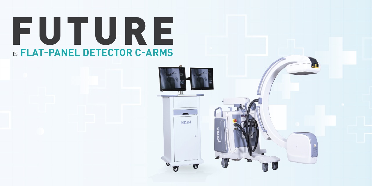
Working as the Surgeon Eye Flat-panel Detector C-arms
Flat-panel detectors have several advantages over image intensifiers, and they are generally expensive compared to image intensifier counterparts. There are several advantages of opting for the FPD C-arm system. This includes:
- FPD C-arms have the potential for higher image resolution and produce a more consistent high-quality digital image.
- FPD C-arms also have some powerful hardware advantages, and investing in them will not make one disappointed.
- FPD C-arms are widely used in interventional radiology for conducting minimally invasive surgery by guiding doctors for accurate positioning during the surgery.
- Their small footprint and low radiation doses, make it to be used flexibly in orthopedics, gynecology, surgery, and other facilities.
- A FPD is shorter compared to an image intensifier because, as it comprises the flat-panel rather than an extended tube structure.
Cost of Flat-panel Detector C-arms
Flat-panel detectors tend to have better image quality and precision imaging capabilities with the recent evolution of technology.
Image intensifiers will usually cost less than flat-panel detector technology. However, the image intensifiers may experience increased downtime due to degradation and on the other hand, flat-panel detectors are advanced technology and become more readily accessible every year. As more options have been introduced, the price of many flat-panel detectors has come down extensively compared to even just a couple of years ago.
Bringing the Best
Trivitron Healthcare Elite/Infinity series of Flat Panel Digital C-Arms
Trivitron Healthcare Elite/Infinity series of Flat Panel Digital C-arms offer robust performance and high precision imaging. An innovative and intuitive surgical C-arm system significantly enhances precision and throughput during surgical procedures along with high precision and high-quality imaging.
It is also equipped with low-dose fluoroscopy made possible with Flat Panel Detector Technology, which makes this C-Arm a perfect addition for a Hybrid OR. It is integrated with tools like PACS and HIS RIS for better access to the data.
Best for Clinical Values
- It comes with a wide range of applications for intraoperative imaging applications in orthopedics, trauma surgery, urology, neurology, gastro, pain management, and peripheral vascular surgery.
- The road mapping feature helps surgeon with ease of working. It guides in finding the path that helps reach the target location in branches during the surgery.
- It comes with DSA or Digital Subtraction Angiography is used in vascular diagnostics. It is very useful for finding blockages or aneurysms in arteries or veins.
If you are looking to purchase a flat-panel detector C-arm system for your facility, experts at Trivitron Healthcare can help you determine the model that is best suited for your needs.


